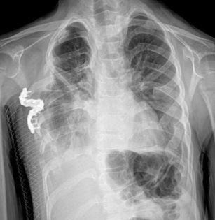
The age of the patient is the most important factor in the treatment plan of the rib cage deformity.
While non-surgical treatments are generally preferred in patients up to 11 years of age, non-surgical or surgical treatment option can be preffered to patients aged 11-17. At this age group (11-17) treatment option determined in a way that will be decided together with the doctor, the patient and the parents by taking into account the expectations from the result of treatment of patient and parents and positive or negative aspects of the each treatment. If you are over the age of 17, generally the surgical treatment method is choosen.
Although age is the most important factor in determining treatment, it is not the only factor. For example, in some deformities under 17 years of age, surgery may be mandatory. Your doctor will recommend the best choice to the patient, taking into account factors such as the flexibility of the rib cage, the shape of the deformity, and possible adhesions. In addition, you will present the possible alternatives to the patient by presenting their positive and negative aspects, and you will make the final decision jointly with the patient and his family, and your doctor.
As Dr. Pectus, we believe that the first option in any case should be non-surgical treatment. Therefore, if applicable, our first choice recommendation is always non-surgical treatments.
Real Treatments
Non-Surgical Treatments
As Dr. Pectus, we believe that the first option in any case should be non-surgical treatment. Therefore, if applicable, our first choice recommendation is always non-surgical treatments.”
Vacuum Bell Treatment
Vacuum Bell Treatment is a medical product, also known as the Vacuum Bell. It is an instrument usually in the shape of a bowl made of orthopedic silicone and connected to a hand pump by means of a hose. The female type ones are made in a different design than the oval or round shape in order not to pull the breast tissues of women. It is used in patients with Funnel chest (Pectus excavatum).
The Vacuum Bell is manually attached to the rib cage by taking the hollow part of the chest into the bowl, with the other hand the pump is squeezed several times so that a negative pressure is created inside the bowl and pulls the hollow part of the chest upwards. The vacuum stays attached to the chest for the periods determined by your doctor, and when this time is completed, when the patient opens the tap of the air drainage system, the vacuum leaves the place where it sticks to the patient.
Vacuum Bell Treatment is ineffective in patients 17 years of age and older, since the development of the bones is completed (the epiphyseal plates of the bones are closed) and the bones can no longer be shaped in this way. (Although it seems to correct the deformity at first when used in this patient group, the chest collapses again after a while).
Since the deformity will recur when this treatment is stopped, the tratment should be continued until the patient reaches the age of 17. In this process, your doctor will follow you regularly at 6 month-intervals. In this period, if you prefer to be examined online you will be also able to be followed up through online consultation. Of course, during the treatment period, the frequency of use of vacum will be gradually reduced to be adjusted by your doctor. For example, if 2 hours of daily use is recommended in the first year of treatment, this period can be reduced to 1 hour in the 3rd year, and after 5 years, it can be used every other day.
On the right patient, the ideal product, the right size, the correct positioning of the product on the patient, vacuuming at the appropriate pressure and other information, Dr. Pectus will support you with all its experience and will provide you with assurance by being accessible for all your problems after the treatment.
Since the deformity will recur when this treatment is stopped, it should be continued until the patient reaches the age of 17. In this process, your doctor will follow you for 6 months and you will be able to follow up through online consultation. Of course, during the treatment period, the frequency of use will be gradually reduced to be adjusted by your doctor. For example, if 2 hours of daily use is recommended in the first year of treatment, this period can be reduced to 1 hour in the 3rd year, and after 5 years, it can be used every other day.
Vacuum Bell Treatment


What is "vacuum treatment" among the non-surgical treatments for pectus excavatum?

Result of only 6 weeks vacuum bell treatment

Can't vacuum bell be used under 2 years old?
Orthosis Treatment
Orthosis treatment is a non-surgical treatment method generally used for the treatment of pigeon chest (Pectus Carinatum) and sometimes Rib Flare deformities. It is based on the principle of regular use of a medical device called Orthosis, which will provide a degree of pressure by supporting from the back to correct the deformity in the area where the anterior dislocation is by providing a freedom from the lateral sides without hindering the movements of the chest and breathing.
Orthosis treatment gives 100% results in patients who are at the age where the bone can be shaped (the epiphyseal plates have not closed yet), that is, in patients aged 17 years and under. Considering the social life of the patient, the orthosis should be worn on the patient for a period of 8 to 23 hours per day. The longer this period, the sooner the results are obtained. For example, in a patient who regularly practices 23 hours a day, the deformity improves 100% at the end of 6 weeks. In a patient who applies it for 8 hours, this process may be prolonged.
Orthotic treatment does not impose restrictions on bathing, swimming and other sports. For example, for an individual who does sports for two hours a day, 2 hours are deducted from the 23-hour period, and these two hours are counted as if the patient used the orthosis.
Correct patient selection, ideal pressure setting, correct positioning of the product on the patient and choosing the right size product are of great importance. For example, a large product may restrict the patient's arm movements. Applying the orthosis at high pressure may cause the patient to transform into a more dangerous funnel chest instead of a pigeon's chest. Or it can cause skin sores. Dr. Pectus will support you with all its experience and will provide you with assurance by being accessible for all your problems after the treatment.
Since the deformity will recur when this treatment is stopped, the tratment should be continued until the patient reaches the age of 17. In this process, your doctor will follow you regularly at 6 month-intervals. In this period, if you prefer to be examined online you will be also able to be followed up through online consultation. Of course, during the treatment period, the frequency of use of orthosis will be gradually reduced to be adjusted by your doctor.
Bandage Treatment
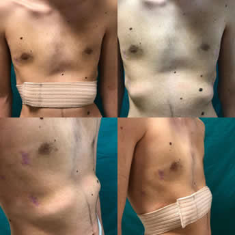
Bandage treatment is based on the principle of using a rib flare bandage, which is similar to an elastic bandage used for rib flare deformity, but corrects the deformity by not losing its elasticity over the years.
For some rib flare deformities, a bandage may not be sufficient, so your doctor may recommend an orthosis. However, it is usually sufficient.
After the bandage is wrapped around the waist in a way that includes the dislocated rib ends, it is tightened to apply pressure to the dislocated ribs and stands on the patient in this way. It remains attached to the patient for certain periods of the day.
Although there is no rib flare in some pigeon chest (pectus carinatum) patients, the rib ends begin to protrude when the orthosis is applied. In such patients, a combined treatment with a protective orthosis can be started in order to prevent rib flare while correcting the pectus carinatum.
Surgical Treatments
As Dr. Pectus Clinic, we always recommend non-surgical treatments as the first option if appropriate for the patient. However, we can apply the entire surgical spectrum of pectus treatment for patients who are not suitable for non-surgical treatment.
These operations are grouped under two headings as “a real treatment surgery” in which the sternum bone is pulled to where it should be, or “correction of the image and psychological effects” by filling the collapsed bone with a silicone implant.
Surgery to Correct the Image and Psychological Effects:
Real Treatment Surgeries:
Minimally Invasive Surgeries
Open Surgeries
MIRPE (Minimally Invasive Repair of Pectus Excavatum) – (Modified) Nuss Procedure
Sandwich Bar
Technique
Surgery to Correct the Image and Psychological Effects
Pectus Excavatum Correction with 3D Silicone Implant
The main purpose of the treatment should of course be a "real treatment surgery", which is the surgery to replace the sternum bone where it should be. However, for real treatment surgery, the patient needs to withdraw from daily life for a month, need someone to take care of him, and struggle with the patient's pain. Moreover, the patient cannot do sports for the first 3 months after the surgery, and then contact sports will be restricted until 3 years after (the bars are removed again). The patient's work-school may not allow him to withdraw from daily life for a month, or he may not have anyone to take care of him. Apart from this, the patient may be an athlete and it may not be possible to stay away from sports for 3 years. There could be other problems, for example, the patient lives abroad and it may be problematic to come and go. For this reason, the patient may not want to undergo a second surgery. Or he might say, "Save me at once, I don't want to deal with this again." Apart from these, they may also be afraid of reasons such as post-operative pain and penetration into the chest cavity.
Our recommendation to patients with all these problems is to hide the deformity by placing a 3D silicone implant specially designed according to the patient's tomography on the collapsed part of the bone. This surgery does not have any of the negative factors of real treatment surgeries that we have written in the above paragraph. However, the only benefit on the patient is that it provides relief from the aesthetic and psychological effects of the deformity.
It is based on the principle of filling the collapsed parts of the thorax with 3D silicone, which is specially produced. After this treatment decision is given to the patient, a 3-dimensional chest tomography is taken, and the images of this tomography are shared with this silicone producing company in France. Then, this silicone implant, which is specially produced for the patient's deformity, is sent to your doctor. After the supply of the implant, the surgery is performed. In the surgery, an incision of approximately 6-7 cm is made on the dimpled part, and this material is placed under the skin and muscles to fill the dimple with the appropriate technique and the incision is sutured. Since fluid will be collected in this area, the injector is dipped daily and the fluid is drained, and the patient is discharged on the 3rd day as this need is also eliminated. It does not need any further surgery. It does not interfere with sports, it does not cause pain. There is no life risk.
This surgery is performed at the age of 17 and above, when the patient completes his development and takes the final shape of the chest cupping.
Poland's Syndrome Treatment with 3D Silicone Implant
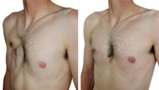
The visual problem arising from the absence of pectoral muscles, which is the main problem in Poland's Syndrome, is eliminated by filling this muscle with a specially produced silicone material.
We recommend this method as the first choice to all patients with Poland's Syndrome, as it is definitely more advantageous to place a silicone implant through a small incision instead of a major surgery such as pulling the muscle from the back to the front and making more incisions in the routinely used method applied by plastic surgeons in Poland's Syndrome.
This surgery is performed in patients with Poland's Syndrome at the age of 17 and above, when they have completed their development.
Minimally Invasive Surgeries
MIRPE (Minimally Invasive Repair of Pectus Excavatum) – (Modified) Nuss Procedure
Minimally invasive repair of Pectus Excavatum is a method used for the treatment of pectus excavatum. MIRPE is based on the principle of lifting the sternum by one or more metal rods that enter the rib cage from the points where the patient's right and left ribs begin to collapse and pass under the deepest point of the sternum bone.
A pediatric surgeon who first described MIRPE, Prof. Dr. Donald Nuss placed this bar with the help of his fingers in young children. Over time, the technique was developed and perfected, and it was named the "Modified Nuss Procedure". In general, all variations are grouped under the MIRPE heading.
As Dr. Pectus, we can perform this surgery in all spectra, including the Cross bar, which can be applied in only a few clinics in the world, and the 4 bar techniques, which we have described for the first time in the world and have been published in the literature in this way. We have the honor of being one of the clinics that performs this surgery the most in the world with our experience in many cases.
Would you like to see how the pectus deformity looks at the heart from the inside with surgical images?
Attention! The video may contain surgical footage. In this regard, we recommend that it not be watched next to sensitive individuals and children.

There is no age limit for this surgery. It can be performed at any age in the appropriate patient. You can reach the article that we published by bringing the largest case series reported in the world at the age of 40 and over to the literature.

Everyone with pectus excavatum deformity should watch it!


Everyone with pectus excavatum deformity should watch it!

Are you curious about the treatment process of our 39-year-old pectus excavatum patient? #DrPectus

Nuss Surgery | Dr. Pectus Clinic

Take an inside look at Nuss surgery
MIRPC (Minimal Invasive Repair of Pectus Carinatum) – Abramson Procedure
Minimal Invasive Repair of Pectus Carinatum is a method developed for the treatment of pectus carinatum. In the operation, no way is entered into the chest cage. It is based on the principle of passing a metal bar horizontally from the right side of the patient to the left side of the patient, under the skin and muscle, but passing over the most protruding point of the sternum bone, and fixing it to the sides of the ribs with the help of wire by pressing. Thus, with the pressure of the bar, the sternum bone is precipitated where it should be.
Since we can provide the same pressure with orthosis without surgery until the age of 17, we usually do not perform these surgeries. There is no upper age limit.
2-4 years after the operation, with a much simpler operation, the bars are removed by entering the previous incisions and the patient is discharged on the same day.
Sandwich Bar Technique
Sandwich Bar Technique is applied in cases where the left or right side of the patient's chest is collapsed and the opposite side is hollow. It is an operation in which both MIRPE and MIRPC are applied. Thus, both pitting and dislocation are eliminated.
2-4 years after the operation, with a much simpler operation, the bars are removed by entering the previous incisions and the patient is discharged on the same day.
Flare Buster Technique
Flare Buster Technique is a technique that we perform during MIRPE or MIRPC surgeries by placing steel wires during surgery in order to remove some rib protrusions, especially rib flare deformity, which had been present in the patient before the surgery or may occur while the surgery.
Flare Buster Technique is applied in conjunction with MIRPE or MIRPC surgeries when deemed necessary by your doctor, and is terminated when the steel wire is removed while the bars are removed.
Open Surgeries
Ravitch Surgery
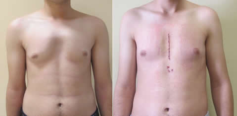
Ravitch Surgery is the oldest and classical thoracic cage deformity correction surgery. It is a type of surgery that can be used in the treatment of both pectus excavatum and carinatum, as well as in pectus arcuatum and asymmetric deformities. While a vertical incision is preferred over the sternum in men, a horizontal incision is made from the folds under the breasts in order to keep the incision scars more hidden in women.
After the incision, some rib cartilages are removed. The sternum is taken to its proper place. Sometimes with this method, materials such as a pectus bar or wire can be placed inside and the bone can be fixed in the appropriate place.
Onen Procedure
Onen Procedure is an open surgery technique that has been brought to the literature by the team that I am also in. One of the first to apply Nuss surgery in Turkey and one of its veterans, my late teacher Dr. We tried to immortalize Ahmet Önen's name by giving his last name to the technique we reported in this article, which we published shortly after his death.
Although this surgery is similar to the Ravitch surgery, it is based on the principle that no cartilage is removed from the patient and instead the sharp part of the bone is partially removed and then fixed with a screw and plate system.
The main place of application is pectus arcuatum. And as one of the developers of this technique, I can say that this is arguably the best treatment for pectus arcuatum.

Cartilage Rib Removal in Rib Flare Treatment
In individuals with protruding rib ends, approximately 3 cm incisions are made over these rib ends, and the membrane feeding the cartilage on the cartilage ribs is opened without tearing, and the cartilage is removed from it. Afterwards, this membrane is sutured end to end in the appropriate position and cartilage tissue formation is provided again. However, this time, the cartilage tissue to be formed will be in a proper position as the membrane is brought into a proper position.
With rib flare surgery, patients achieve a much thinner waist circumference and posture than tiny incisions. As Dr. Pectus Clinic, we do the surgery in a way that cartilage is formed again, as we mentioned above, so there is no organ loss in the patient.
Lateral Thoracic Expansion
Lateral Thoracic Expansion is applied for the treatment of thoracic dystrophies (the most common among them is jeune syndrome) and other small rib cage deformities (for example, when rib anomaly accompanies another component of Poland's Syndrome, other than the absence of classical chest muscle, the rib cage may remain small). With a vertical incision from the side at the armpit level, the rib cage is expanded from the sides by reaching from the 4th rib to the 9th rib and making a connection between the ribs in a different way.
The purpose of this surgery is to widen the thoracic cage, making room for the patient's heart and lungs to grow, and thereby enabling the patient to continue his or her life. By applying the same operation to the opposite side within 6 months or 1 year, asymmetry is prevented and the lung on the opposite side can find a place to expand.
In this surgery, since we do not leave the expansion to screws and plates, and we apply it by combining bones in different formations, bone formation can continue over the years as the patient grows, and the thorax can expand as well, and strength is ensured by refilling that area with the bone tissues that the patient will create in that area. Thus, various complications do not occur with the dislocation of foreign objects such as screws and plates.
Dynamic Thoracoplasty
Dynamic Thoracoplasty is applied for the treatment of thoracic dystrophies (the most common among them is jeune syndrome) and other small rib cage deformities (for example, when rib anomaly accompanies another component of Poland's Syndrome, other than the absence of classical chest muscle, the rib cage may remain small). Although lateral thoracic expansion (see) is applied from both sides of the patient as the first choice in these patients, lateral expansion may be insufficient with the rapid development of the patient. In this case, expansion can be provided from the front as well.
Similar to open heart surgery, the sternum bone is split vertically in half. The pectoral muscle (major pectoral muscle) on both sides is extended towards this area and placed between the two parts of the sternum bone. Since there is a muscle tissue fed between the two sternum pieces, the sternum pieces are prevented from growing towards each other and sticking again. As the patient grows, the rib cage may also expand.
The aim of this surgery is to widen the thoracic cage, just like in lateral thoracic expansion, to make room for the patient's heart and lungs to grow, and to enable the patient to continue his/her life.
Since the patient's own muscle tissue is extended between the bone in this surgery, there is no foreign material or related complications. As the patient grows, the muscles will also grow, helping to stretch the bone and expand the rib cage. That's why it's called "dynamic", not fixed.

Sternal Cleft Repair
The sternum bone consists of two vertical leaves while in the womb. While in the mother's womb, these leaves combine to become a single bone. However, since this union does not occur in cleft cases, the heart and internal organs remain unprotected and cause a bad appearance. Sometimes there may be no skin integrity along with the bone. In this case, the heart may be beating outside the patient.
Sternal cleft treatment is surgically bringing the bones end to end and suturing them together in this way as soon as possible. Thus, fusion is achieved at an early age, and bone integrity is ensured before the heart and internal organs grow and become compressed. In the future, after the bone is fused, it grows and develops as in a normal individual and provides room for the internal organs to grow.
However, in case of delay, the bones may not come together as the internal organs grow and develop. For this reason, it may be necessary to put parts taken from different bones of the patient or synthetic materials.
Contact Us
If you have any questions about treatments or would like to schedule an appointment, please fill out the contact form or call +90 501 134 40 88 or email to info@drpectus.com.


