Pectus Excavatum (PE) deformity, also known as Funnel Chest, is characterized by the collapse of the front part of the sternum and chest wall into the chest cavity. It is the most common of all chest wall deformities, with a frequency of 0.4% in the population, and is five times more common in men.

But what are the causes of this deformity? When we research this issue in the scientific literature, we see that no clear reason has been revealed yet. However, some hypotheses have been put forward to try to explain it. Let’s look at these hypotheses together:
Why Does Pectus Excavatum Occur? Pectus Excavatum Associated Conditions
1) Is Pectus Excavatum Genetic
The etiology of pectus excavatum remains a subject of ongoing investigation within the medical community. While geneticists have not definitively established its genetic nature, there is growing evidence suggesting a familial predisposition to this condition. For instance, studies have indicated that 40% of children exhibiting these deformities have relatives with similar rib cage abnormalities. This familial occurrence raises questions about the genetic component of pectus excavatum.

Despite the absence of clear categorization as a genetic disorder in current literature, recent research has unveiled potential genetic markers associated with this condition. This emerging data hints at genetic involvement, although it has yet to be fully recognized as such within established medical literature. Therefore, while pectus excavatum is not explicitly labeled as a genetic disorder, ongoing studies are shedding light on its potential genetic underpinnings.

In light of these findings, the question arises: “Is pectus excavatum a genetic disorder?” While not definitively confirmed, the accumulating evidence of familial predisposition and emerging genetic markers prompts further exploration into the genetic aspects of this condition.
This integrates the question about the genetic nature of pectus excavatum into the discussion about ongoing research and the familial tendencies observed, leading to a logical flow within your article.

2) Intrauterine Pressure
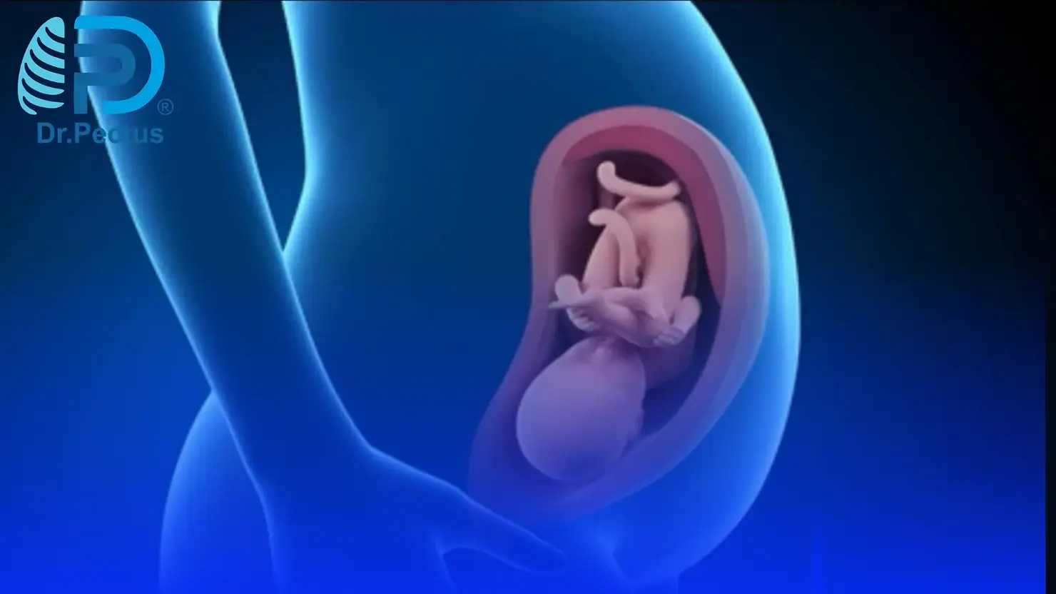
Intrauterine Pressure is stated in the literature that any person’s knee or elbow that puts pressure on the breastbone of the fetus while it is in the womb may cause pectus excavatum.

3) Rickets: Signs in the Chest
Rickets, a prevalent bone disease, arises from a deficiency in vitamin D within the body. Vitamin D plays a crucial role in bone development, and its deficiency can lead to various bone-related issues, including sternum complications. The sternum, like other bones in the body, may be affected when vitamin D levels are insufficient. One notable manifestation of such deficiency is the development of pectus excavatum.

What are the signs of rickets in the chest?
Pectus excavatum, characterized by the inward depression of the sternum, is among the significant indications of rickets affecting the chest area. This condition, resulting from inadequate vitamin D, underscores the importance of this vitamin in maintaining proper bone structure, particularly in the chest region. The appearance of pectus excavatum can be one of the notable signs suggesting rickets in the chest, highlighting the link between vitamin D deficiency and bone deformities in this area.
4) Restrictive Lung Diseases
Impact of Pectus Excavatum on Breathing and Lung Capacity
Among the various conditions affecting lung functionality, restrictive lung diseases stand out. These diseases entail a loss of flexibility in the lungs, hindering their ability to fully expand upon inhalation. Consequently, the volume of air the lungs can accommodate diminishes.

One of the notable occurrences reported in the literature links the development of pectus excavatum to the reduction of intrathoracic pressure during early childhood. This reduction in pressure can lead to the inward collapse of the sternum bone, resulting in pectus excavatum.
Moreover, the presence of pectus excavatum itself has been observed to exert pressure on the lungs, consequently contributing to the development or exacerbation of restrictive lung diseases.

Does pectus excavatum affect breathing and lung capacity?
Pectus excavatum can significantly impact breathing due to its compressive effect on the lungs. The inward indentation of the sternum caused by pectus excavatum may restrict the normal expansion of the lungs during inhalation, potentially leading to breathing difficulties.
Furthermore, this condition can impede lung capacity, limiting the volume of air the lungs can comfortably hold.
The presence of pectus excavatum poses a dual challenge—first by its potential role in causing restrictive lung diseases through its impact on intrathoracic pressure, and second by directly compressing the lungs, further compromising respiratory function and lung capacity.
5) Aperture (Diaphragm) Anomalies
In the literature, it has been stated that when the diaphragm, which is the muscle that separates the chest and abdominal cavities, is congenitally short in a patient, this situation can cause pectus excavatum by collaps the sternum bone.

Other studies have shown that abnormal contraction of the diaphragm due to some problems in the neuromuscular junction may also cause this condition. In addition, according to other hypotheses, in case of strong chest muscles and weak diaphragm, the sternum turns outward and pectus carinatum occurs, and in case of weak chest muscles and strong diaphragm muscle, the sternum collapses inward and pectus excavatum occurs.
6) Internal Diseases During the Production Phase of Bones and Cartilages
The ribs that form the anterior chest wall extend approximately 2-3 cm next to the sternum bone. Cartilages provide the 2-3 cm remaining connection between them. If there are problems with the production of these bones and cartilages in the body during the womb or later development period, various deformities, especially pectus excavatum, may develop due to the impaired production of these bones or cartilages.

7) Defects in the Structure of Type 2 Collagen, a Component of Rib Cartilage
Diseases characterized by deterioration in the structure of Type 2 collagen, which is generally the filling material of cartilage tissue, may deteriorate the cartilage structure and lead to collapse of the sternum and may be associated with pectus excavatum.

8) Abnormal Levels of Zinc, Magnesium or Calcium in the Blood
Scientific studies show that cartilage rib problems and related funnel chest may develop in patients with decreased zinc levels and increased calcium and magnesium levels.

9) Some syndromes present in the patient:
a. Achondroplasia and Pectus Excavatum
Achondroplasia is a genetically inherited cartilage development disorder. 35-53% of patients have volume reduction in the rib cage. Chest circumference has decreased by 25-30% compared to individuals of average height. Especially in men, while the horizontal diameter is closer to normal, the anterior-posterior diameter becomes considerably smaller.

b. Poland Syndrome and Pectus Excavatum
Poland syndrome is a syndrome manifested by the absence of the chest muscle (pectoral muscle). In almost all patients, one side (mostly the right half of the body) is affected. Poland syndrome is not a genetic disease. Familial transmission is less than 1%.

The reason has not clearly revealed. Other components of the syndrome include the absence of also some other muscles in the body other than the pectoral muscle, breast and nipple abnormalities or their absence, underdevelopment or absence of structures in the ribs and chest wall, absence of axillary hair growth, presence of various anomalies or deficiencies in the arms and/or hands on the affected side.
One or many of these other components may or may not accompany the absence of pectoral muscle. Treatment is planned separately for each component. Pectus excavatum can be observed as a component that can be classified as chest wall deformities in patients with Poland’s Syndrome.
c. Anterior Thoracic Dysplasia and Pectus Excavatum
As in Poland syndrome, there are some problems with underdevelopment or absence of the breast on the affected side and underdevelopment or absence of structures in the chest wall. Unlike Poland syndrome, pectoral muscle absence is not observed.

d.Congenital Rib Anomalies and Pectus Excavatum
Some problems in rib development that can result in short ribs or intrathoracic ribs can also cause pectus excavatum.

e. Thoracic Dystrophies and Pectus Excavatum
Thoracic Dystrophies is a condition where the rib cage remains small. Thoracic Dystrophies can affect many organs. It is genetically inherited. There are 22 varieties. The most common one is asphyxiated thoracic dystrophy, or “Jeune Syndrome”.

Other well-known ones; Saldino-Noonan Syndrome, Majewski Syndrome, Mainzer-Saldino Syndrome, Beemer-Langer Syndrome, Ellis-van Creveld Syndrome. The common findings of all phenotypes are inadequate development of the rib cage (thoracic hypoplasia) and short ribs. These can lead to various chest wall deformities, especially pectus excavatum.
f. Noonan Syndrome and Pectus Excavatum
Noonan Syndrome is characterized by short stature and congenital heart disease. Noonan Syndrome is genetically inherited. Noonan Syndrome is seen equally in men and women. It has components that affect many organs. The skeletal component includes chest wall deformities and pectus excavatum.

g. Marfan Syndrome and Pectus Excavatum
Marfan Syndrome is a genetic disease. While it can manifest itself with its individual components, it can also have so many components that it can cause problems starting from the neonatal period. Marfan syndrome most commonly manifests itself with eye, heart and musculoskeletal system anomalies.

However, Marfan syndrome can also affect other organs. Rib cage deformities are also seen among skeletal anomalies. Pectus excavatum is worth one point in the criteria developed to diagnose Marfan syndrome.

h. Osteogenesis Imperfecta and Pectus Excavatum (Sunken Chest)
This genetic condition is also known as “glass bone disease” among the public. Osteogenesis imperfecta is related to the fact that bones can break very easily in these patients. Type 1 is associated with a problem in collagen production. It has also been reported in the literature to affect the chest wall bones, although it is rare.
i. Spinal Muscular Atrophy (SMA) and Pectus Excavatum (Sunken Chest)
Spinal muscular atrophy (SMA), a genetic disorder impacting the nervous system and causing progressive muscle weakness, is known for its rare occurrence. However, an intriguing association between SMA and pectus excavatum, commonly referred to as a sunken chest, has been observed in affected individuals.
In SMA, particularly among SMA type 1 patients and some SMA type 2 patients, the uncontrolled contraction of the diaphragm muscle can result in an imbalance in the mechanics of breathing. This imbalance can lead to an inward collapse of the rib cage during breathing, rather than the expected outward expansion. Over time, if left untreated or unmanaged, this altered breathing pattern can contribute to the development of pectus excavatum.
The significance of timely intervention becomes apparent in preventing the onset of pectus excavatum in SMA patients. Early treatment and management strategies that address the breathing mechanics and associated muscular imbalances may help mitigate the risk of rib cage collapse and the subsequent development of pectus excavatum in these individuals.
Understanding this link between SMA and pectus excavatum highlights the complex interplay between muscular disorders and skeletal deformities, emphasizing the importance of comprehensive care and tailored interventions to address both the underlying condition and its potential secondary complications.
Therefore, while SMA primarily affects muscle movements due to nervous system impairment, its connection with pectus excavatum underscores the need for proactive measures to manage respiratory complications and potential skeletal deformities in affected individuals.
j. Loey-Dietz Syndrome (LDZ) and Pectus Excavatum (Sunken Chest)
Loey-Dietz Syndrome (LDZ) is a genetic syndrome characterized by vascular problems (ballooning and ruptures in the vessels called aneurysms), skeletal findings (pectus excavatum is the most common), some features on the head and face (widely spaced eyes, strabismus, cleft palate, premature closure of the fontanelles in the skull) and presence of some skin findings (velvety and translucent skin, easy bruising and unusual scarring). The biggest problem is ballooning in the veins. Therefore, it requires frequent monitoring.
k. Ehler Danlos Syndrome and Pectus Excavatum (Sunken Chest)
Ehler Danlos is a hereditary disease. Ehler danlos is caused by a structural disorder in the body’s filling tissues, called connective tissue. Looseness in tissues is at the forefront. Therefore, it can cause problems with the skin, joints and blood vessels. Extreme flexibility is typical. Pectus excavatum can often be seen as a component of this syndrome.
l. Celiac Disease and Pectus Excavatum (Sunken Chest)
Celiac disease is basically a disorder that occurs as a result of an allergic reaction to the protein called “Gluten” found in grains such as wheat, barley and rye, and as a result, some changes occur in the initial part of the small intestine.
Accordingly, the absorption of some food components such as iron and folic acid from the intestine to the blood is impaired and additional problems may arise due to their deficiencies. It has been reported that familial transmission is common. It has been reported that approximately 1% of celiac patients are accompanied by pectus excavatum for an unexplained reason.
m. Charcot-Marie Tooth Disease and Pectus Excavatum (Sunken Chest)
Charcot-Marie Tooth Disease is a disease related to the nervous system. After damage to the nerves outside the brain and spinal cord, weakening of the hand and foot muscles is observed. One of the components of this syndrome is pectus excavatum or other chest wall deformity, and this component may also accompany the disease.
n. Atelosteogenesis Syndromes and Pectus Excavatum (Sunken Chest)
Atelosteogenesis Syndromes are a syndrome with various types. Their common feature is the presence of problems with the development of bones in the body. Affected individuals are born with feet that turn inward and upward (clubfeet) and hip, knee, and elbow dislocations.
Bones in the spine, ribcage, pelvis, and limbs may be underdeveloped or, in some cases, absent. As a result of limb bone abnormalities, individuals with this condition have very short arms and legs. As the sternum bone is affected along with all bones, various chest wall deformities and pectus excavatum may be accompanied.
o. Pierre-Robin Syndrome and Pectus Excavatum (Sunken Chest)
The syndrome basically has three components. These; It is the presence of cleft palate as a result of inadequate development of the lower jaw, the tongue being pulled back and the palatal bones not fusing together.
Pectus excavatum, rib anomalies and inadequate development of the lower ends of the scapula are other common components.
p. Sprengel Deformity and Pectus Excavatum (Sunken Chest)
Sprengel Deformity is a congenital condition that one-sided shoulder blade (scapula) remains high. Sprengel deformitycan often be accompanied by other bone pathologies. Rib cage deformities and pectus excavatum are among the most common.
q. Sotos Syndrome and Pectus Excavatum (Sunken Chest)
Sotos Syndrome is a syndrome characterized by excessive growth of bones. His birth weight and height are high. The arms, legs, hands and feet are large. A long narrow face and redness on the cheeks are typical.
The skull is extremely large. Apart from this, cardiovascular, urinary tract and spine anomalies are common. The brain is underdeveloped and may be accompanied by learning disabilities and mental retardation. It may also be accompanied by chest wall deformities due to excessive growth in the bones.
r. Radial Club Syndrome and Holt Oram Syndrome Radial Club Syndrome and Holt Oram Syndrome
Radial Club Syndrome is characterized by underdevelopment of the Radius bone, which is the main bone that forms the forearm. Bending of the hand from the wrist towards the thumb is also seen.
The thumb may be absent or underdeveloped. This condition can occur alone or be a part of various genetic syndromes. It has been reported in the literature that pectus excavatum may accompany the radial club as a part of Holt-Oram Syndrome.
s. Jarcho-Levin Syndrome and Pectus Excavatum (Sunken Chest)
There is a problem in the junction of the ribs with the spine. Some ribs has fused together. The appearance of the rib cage resembles a “crab” shape.
Nervous system disorders may also occur due to spinal anomalies. Due to rib anomalies, chest wall deformities and pectus excavatum may be observed.


